SBPMD Histology Laboratory Manual
The Eye: Micrograph
Examine the electron Micrographs so that you understand the ultrastructural equivalents of the structures you have seen under the microscope.
Retinal Pigment Epithelium (human) | |
| Click to see enlarged view | |
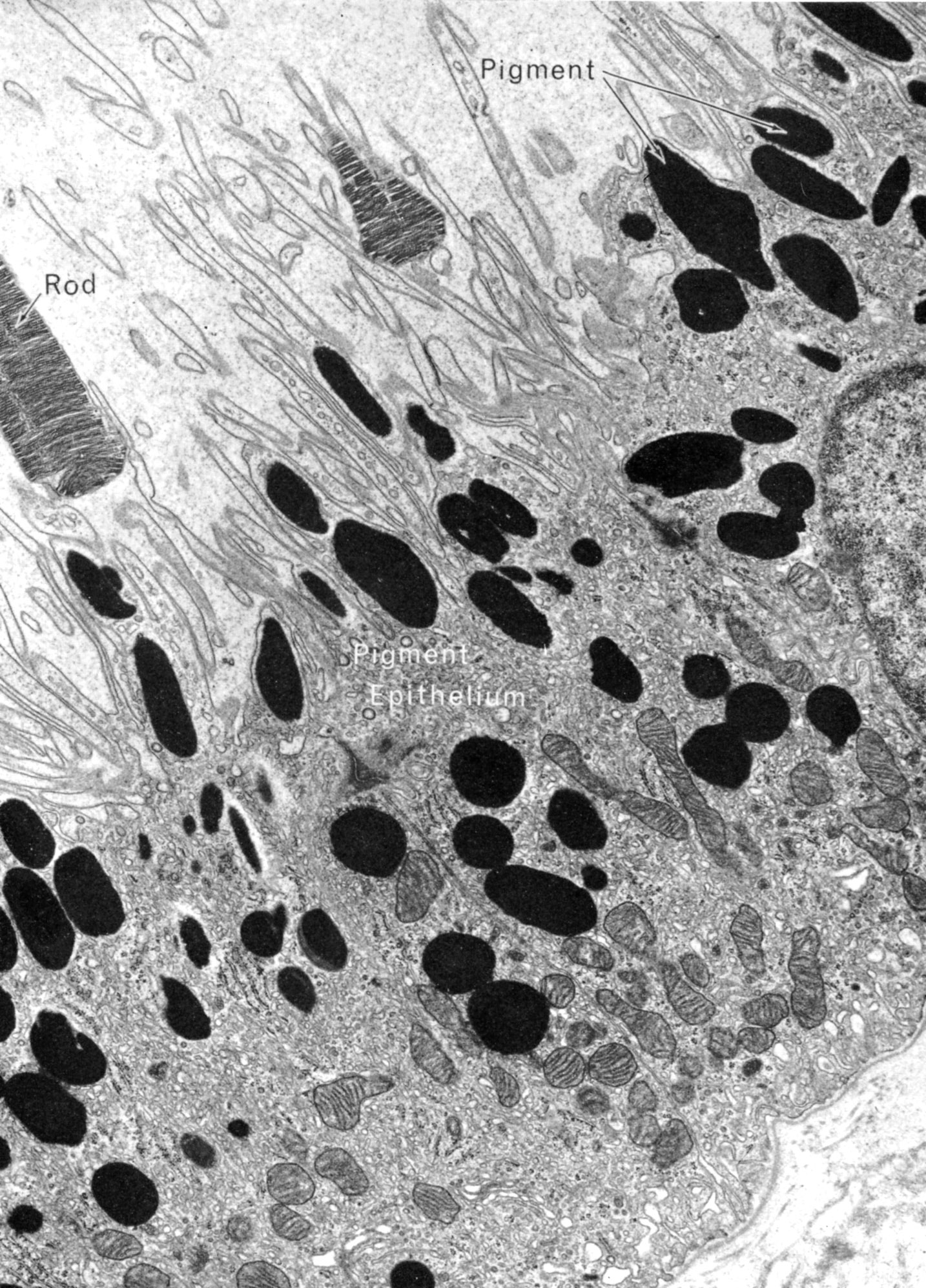 |
Retinal pigment epithelium is made up of cuboidal cells that rest on Bruch’s membrane (lower right). Their apices interface with the outer segments of the rod and cone cells. They absorb excess light, phagocytose the outer segment of photoreceptor cells and participate in the resynthesis of visual pigment. Their lateral borders are connected by junctional complexes consisting of gap junctions and elaborate zonulae occludentes and adherentes which are a component of the blood-retina barrier. Fawcett DW, The Cell: An Atlas of Fine Structure, WB Saunders, Philadelphia, 1966, p. 289.
|
| |