SBPMD Histology Laboratory Manual
Ovary
#61 Ovary, Adult Monkey.
Open with WebViewer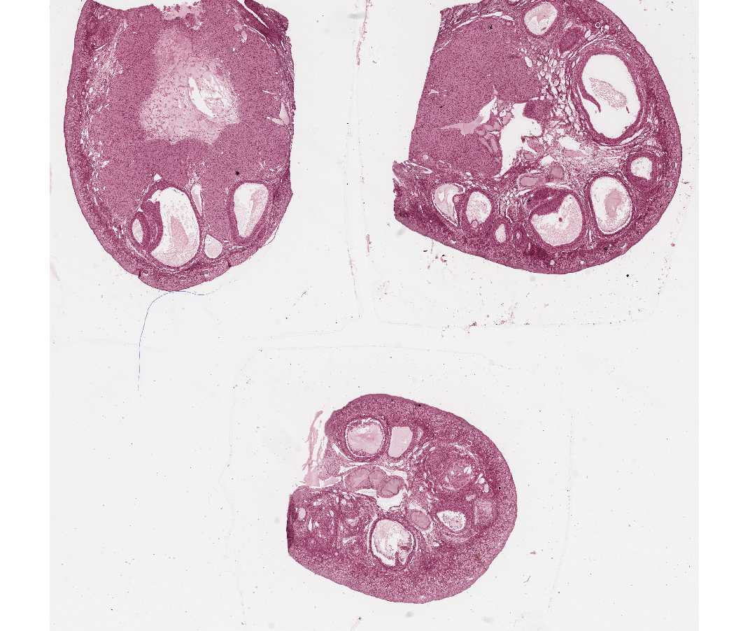
Note: There are 3 sections of ovary on this slide. Examine with the scanning objective, and note the general division of the ovary into an outer cortex (containing follicles in various stages of development) and an inner medulla (containing numerous blood vessels and dense fibrous connective tissue).
Identify:
- Lining epithelium (classically called germinal epithelium), a simple cuboidal covering the ovary, continuous with the mesothelium of the peritoneum.
- Primordial follicles - 1o oocytes surrounded by a single layer of squamous granulosa cells. This is a resting stage.
- Primary follicle - 1o oocyte surrounded by cuboidal or columnar granulosa cells, first as a single layer, and then multi-layered. Theca forms outside this. Zona pellucida surrounds oocyte. These are growing follicles
Secondary or antral follicles 1o oocyte surrounded by granulosa cells among which fluid-filled spaces are coalescing into a single space, or antrum. Outside the basal lamina of the granulosa layer the theca has differentiated into a theca interna and a theca externa. - Mature preovulatory or Graafian follicle - characterized by a very large, central antrum. Zona pellucida is very conspicuous. 1o oocyte is surrounded by a layer of granulosa cells (the corona radiata) and rests on a small mound of granulosa cells called cumulus oophorus.
- Atretic follicles - Note that follicles may undergo atresia during any stage of development. Atresia is often first recognized in the granulosa cells; the nuclei become apoptotic and there is a loosening of the cells. The underlying basement membrane thickens and is known as the glassy membrane. The collapse of the glassy membrane in large follicles that become atretic forms a remnant with the appearance of a rubber band. The theca interna of atretic follicles generally hypertrophies and proliferates to assume a more glandular appearance. This altered theca interna of atretic follicles is then called interstitial gland tissue. The function or importance of this tissue in the human is unknown, but it may occupy a large portion of the ovary in many rodents, rabbits and carnivores.
- Corpus luteum A corpus luteum is the follicular cells (both granulosa and luteal) of an ovulated follicle that have been luteinized. These form a large, steroid-producing organ.
- Corpus albicans This is the connective tissue scar remaining from a degenerated corpus luteum. These are more easily identified in the trichrome slide (following).
#62, #63 Ovary, Adult Human (Mallory's trichrome or H&E) (#62 not scanned)
Open with WebViewer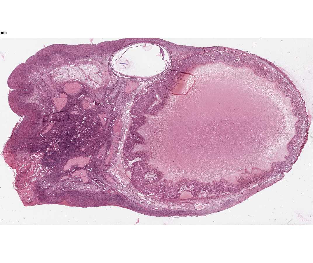
With the naked eye, one can easily distinguish the follicles. Under the microscope, note the scarcity of primary follicles, suggesting that this ovary is from an older woman. Many larger follicles are in various stages of atresia. (The basement membrane that separates the granulosa cells from the vascularized theca interna persists in these atretic follicles and appears as a collapsed structure the glassy membrane.) Identify a corpus albicans (the connective tissue scar remaining from a degenerated corpus luteum, albicans, pl. corpora albicantia).
Some ovary sections contain a recently formed, corpus luteum that dominates the ovary (can be seen with the naked eye). Notice the folding of its wall and the large central cavity filled with coagulum. With the microscope identify the two primary cellular components of the corpus luteum: the granulosa lutein and theca lutein cells. Notice the relationships of these two cell types to each other and to the vascularization of the developing corpus luteum. The theca lutein cells follow the pathways of the invading blood vessels.
#64 Ovary, Adult Human, Corpus Luteum of Pregnancy
Open with WebViewer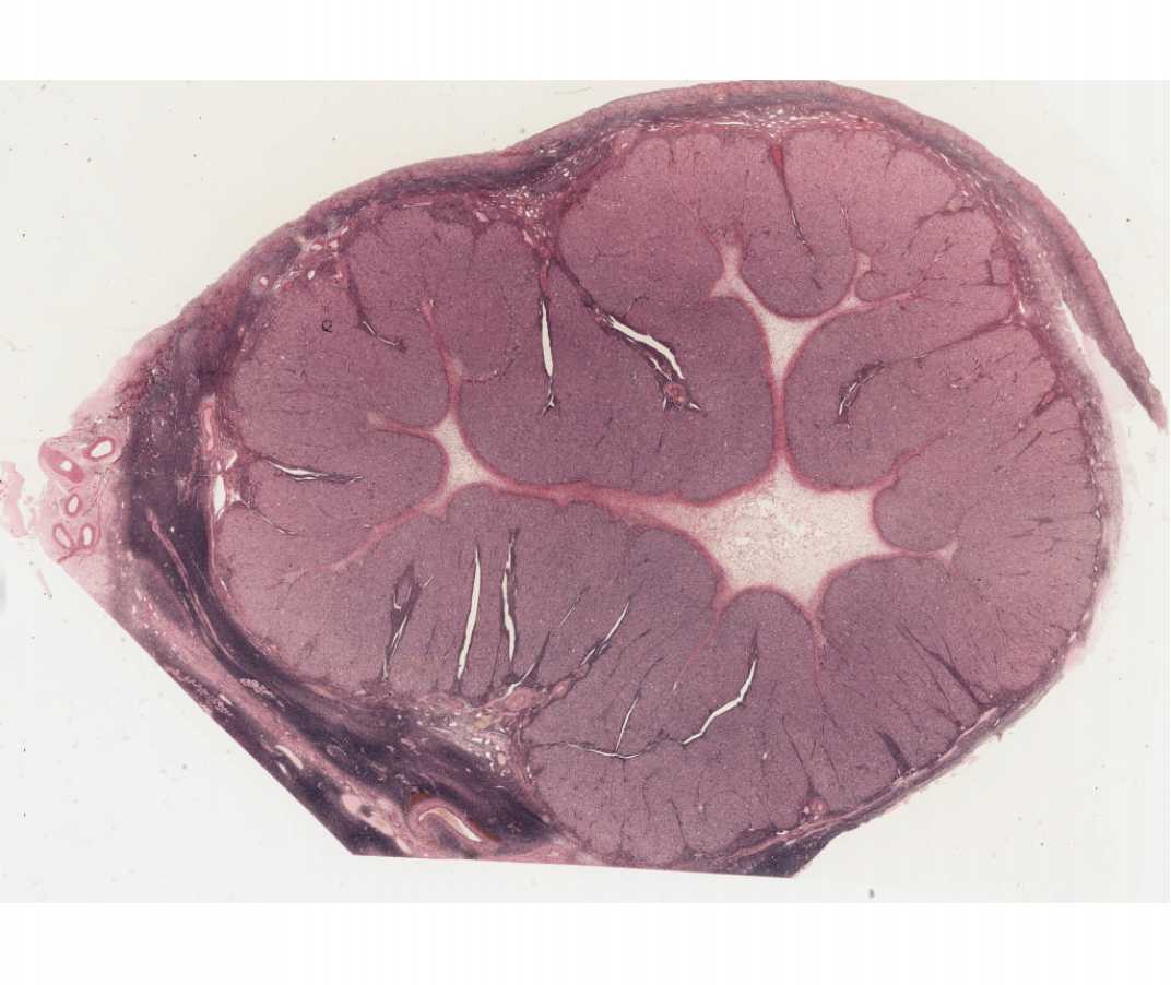
Compare the development of this corpus luteum of pregnancy (#64) (probably from the first trimester) with that of the recently formed corpus luteum of slide #63. Note particularly the increase in thickness of the granulosa luteal (luteal and lutein are equivalent terms) layer, as compared to the thin, peripheral zone of theca luteal cells. The extensive vacuolization of the granulosa luteal cells is due to the extraction of its abundant lipid droplets. This reflects the importance of the corpus luteum (particularly the granulosa lutein cells) as the primary ovarian source of the steroid hormone progesterone.
What is the primary ovarian source of estrogenic hormones?
What are the fine structural specializations of ovarian cells involved with the production of steroid hormones?
Be certain that you understand the changes that occur within the follicle during follicular development.
Consider the hormones of the anterior pituitary involved in follicular growth and ovulation.