| [an error occurred while processing this directive] | |||||
Design of Micro-Surgical Manipulators for Dual-arm Microsurgery
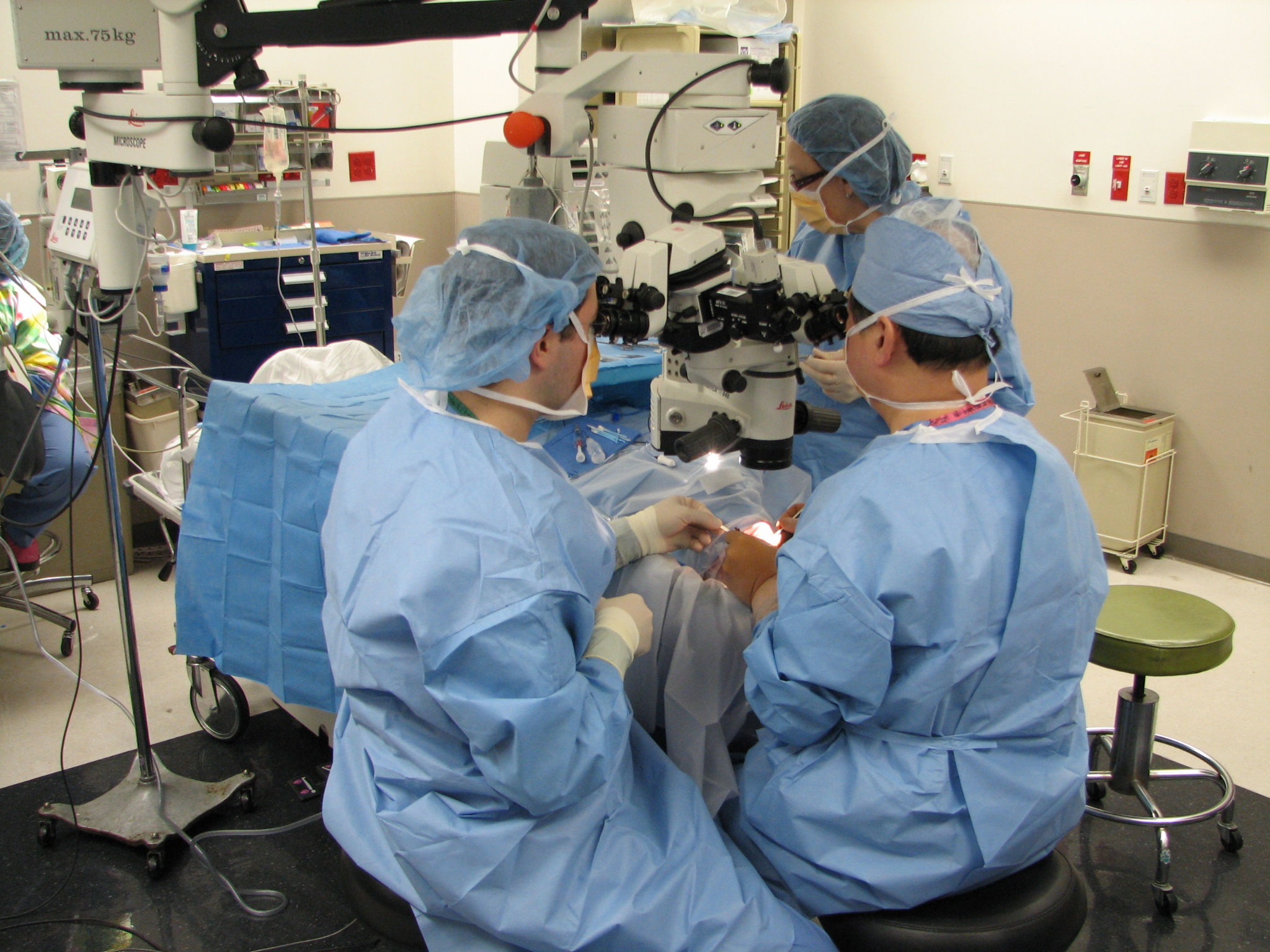
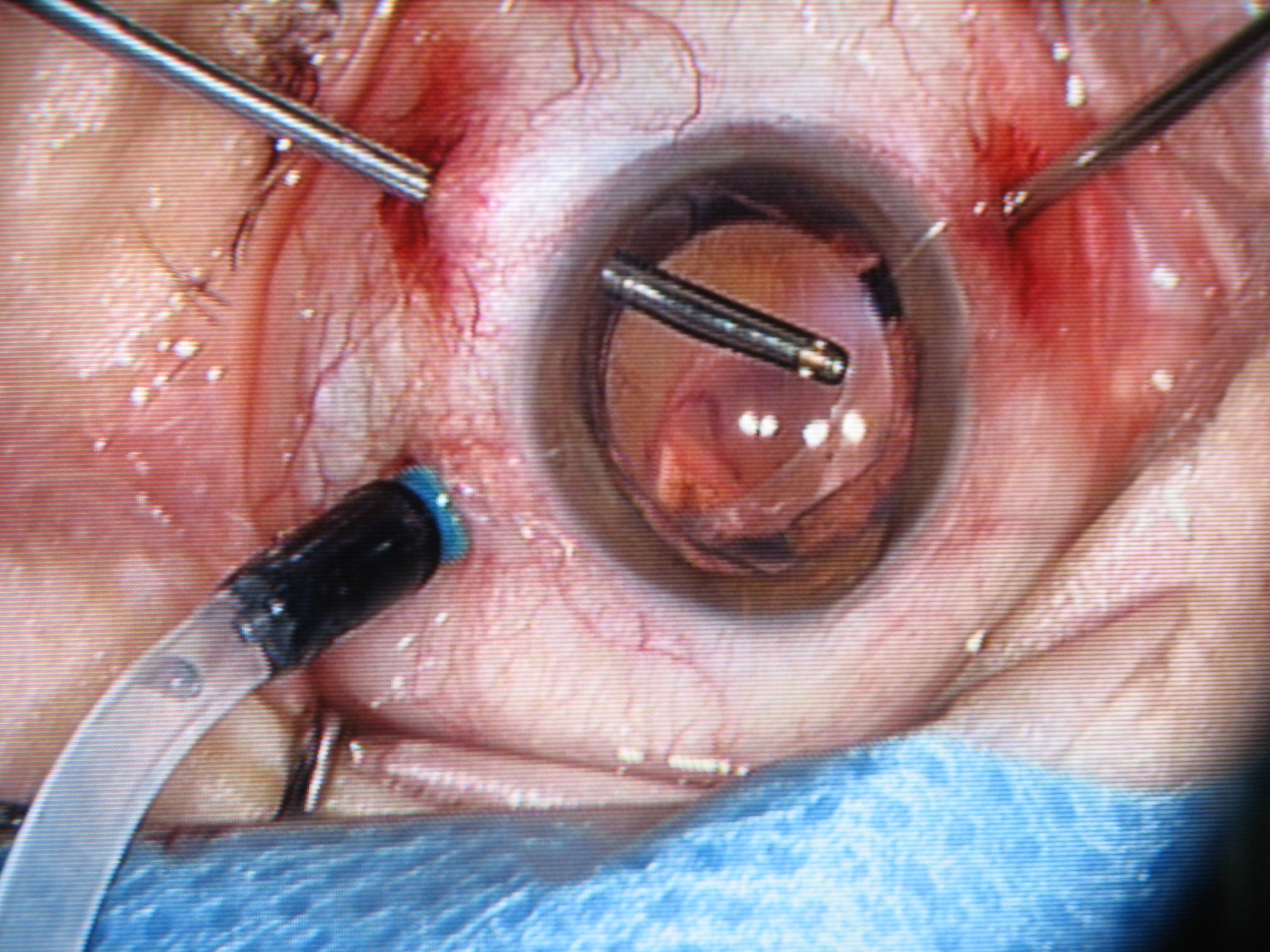
An Overview of the Ophthalmic Surgery Setup In an ophthalmic surgery setup, two surgeons coordinate activities to perform the ophthalmic surgical procedures. The main surgeon sits superior to the patient'ss head and performs most of the surgical tasks including manipulation of the surgical tools and the light source. The assistant surgeon sits beside the patient's head to provide irrigation and removal of fluids and to adjust the placement of the visualization of the external lenses. There is also another assistant sitting on the other side of the patient for tool delivery to the main surgeon. Three incisions are typically made in the sclera to provide access to the inner vitreous body for one irrigation tube, one light source and one surgical instrument. The irrigation tube injects liquid to maintain intraocular pressure. The light source illuminates an area on the retina for proper visualization under the microscope. The surgical tools including picks, micro tweezers, vitrectomy cutters and other laser ablation devices vary dependent on the requirements of the procedure. The surgeons operate using a microscope while visualizing the retina through a dilated iris. Because the visual field does not include the entire retinal surface, the surgery often requires to tilt the eye under the microscope in order to view peripheral areas of the retina.
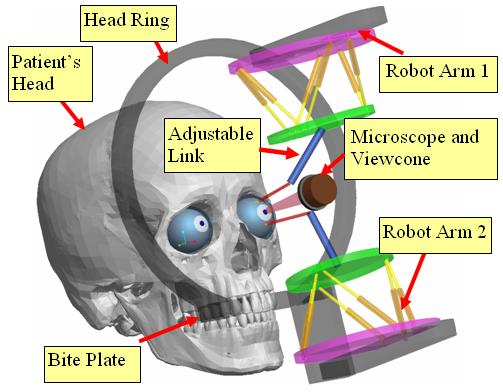
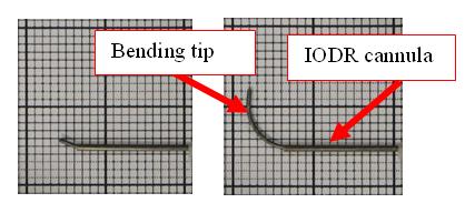
Robot Setup for an Ophthalmic Surgery Our role in this project is to design a 16-DoF hybrid robotic system to assist ophthalmic surgery as shown in the above figure. This system will allow high precision dexterous manipulation of surgical instruments within the vitreous body of the eye and macroscopic rotation of the eye for surgical field visibility. A frame is secured without trauma to the patient's head with a locking bite-plate and coronal strap that rigidly affixes the frame to the patient. Two identical hybrid robotic arms are rigidly attached to this frame. Each robotic arm is composed from a 2 DoF IODR (Intra-Ocular Dexterity Robot) and a 6 DoF parallel robot. This design will allow 5 DoFs for the surgical tools inside the eye, as compared to 4 DoF tools used currently. The 2 DoF IODR provides intra-ocular dexterity while the parallel robot provides global precise positioning of the eye and the surgical tool inside the eye. The IODR utilizes a pre-shaped NiTi tube that bends in one DoF as it is extended outside of a straight cannula. The cannula of the IODR rotates about its axis to provide the second DoF inside the eye. For the parallel robot, we propose the Stewart-Gough platform design due to its rigidity, compactness, positional accuracy, and high payload-to-weight ratio.
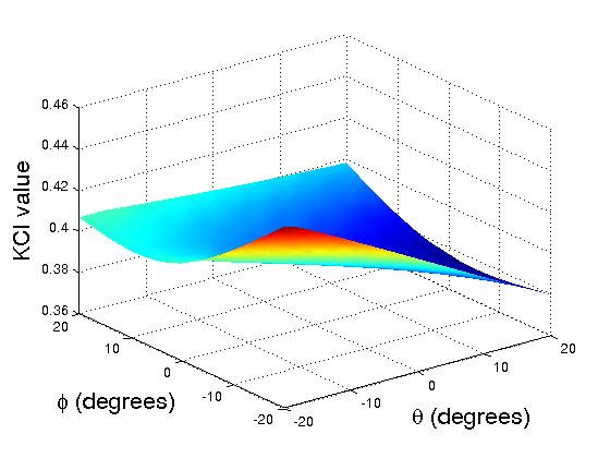
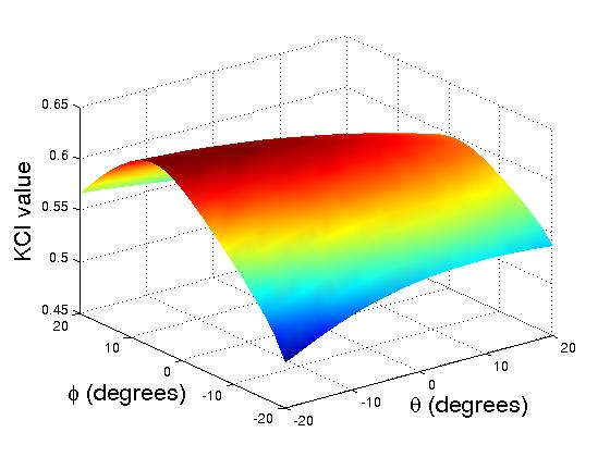
The 2-DoF Intro_Ocular Dexterity Robot An initial performance evaluation is implemented to compare the dexterity between the robotic arms without distal dexterity and with distal dexterity. We plot the KCI (Kinematic Conditioning Index) measurement for both translational and rotational motions. Translational KCI Plots for One Arm without Distal Dexterity (left) and with Distal Dexterity (right)
Rotational KCI Plots for One Arm without Distal Dexterity (left) and with Distal Dexterity (right) Publications:
Collaborators: Dr. Stanley Chang & Dr. Howard Fine Department of Ophthalmology, Columbia University. |
|||||
| [an error occurred while processing this directive] | |||||